


Meet Our Experts

Our team of dedicated access representatives are here to connect you with the specialists you need.
Innovative Research

Our researchers are helping to turn the discoveries we make into tomorrow's advanced treatments for our patients.
Patient and Caregiver Support
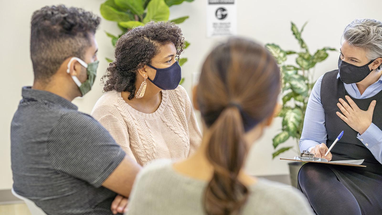
We treat the whole person, not just the cancer. Our support services provide care and support outside of traditional medical care.
News
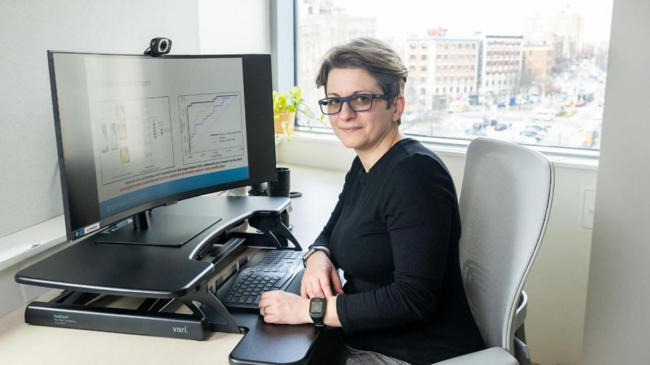
- April 1, 2024
Despina Kontos, PhD, shares how artificial intelligence in imaging is revolutionizing cancer care.
Topic
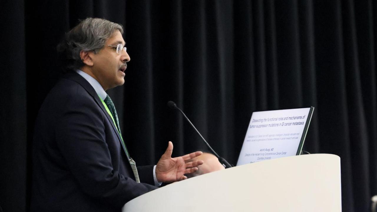
- April 2, 2024
Dozens of HICCC researchers will present their research at this year's AACR annual meeting.
Topic
-
Source:
CUIMC NewsroomApril 8, 2024A new type of investigational therapeutic for pancreatic cancer has shown unprecedented tumor-fighting abilities in preclinical models of the disease.
Topic
-
Source:
Cancer DynamicsMarch 21, 2024IICD Researchers introduce an innovative computational tool named Starfysh, designed to revolutionize the study of gene expression within tissues.
Topic
- February 1, 2024
The 2023 HICCC Annual Report is live. Read more about how we're working to end cancer for good.


Patient Stories
-
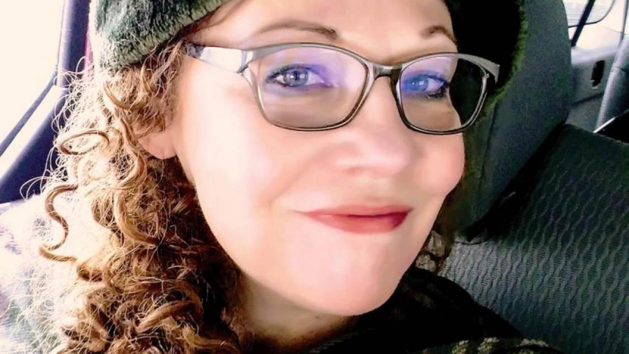
After Patti Murillo-Casa beat cervical cancer, she had another fight for her health - depression led to weight gain and a series of other health issues. After losing 113 pounds, Patti is a group fitness instructor specializing in senior fitness and cancer recovery fitness, sharing her story with others to inspire change.
-
Jane Hanley was devastated when she learned she had advanced lung cancer. Nearly five years later, Jane has a new outlook on life under the care and trust of her devoted oncology team at Columbia/NewYork-Presbyterian.

Events
-
- Tuesday, May 7, 20244:00 PM to 5:00 PM
Ways to Give

When you give to the HICCC, you are making a difference in the lives of cancer patients. Your support accelerates the discoveries made in our labs into the innovative cancer care of tomorrow.